
Experiment
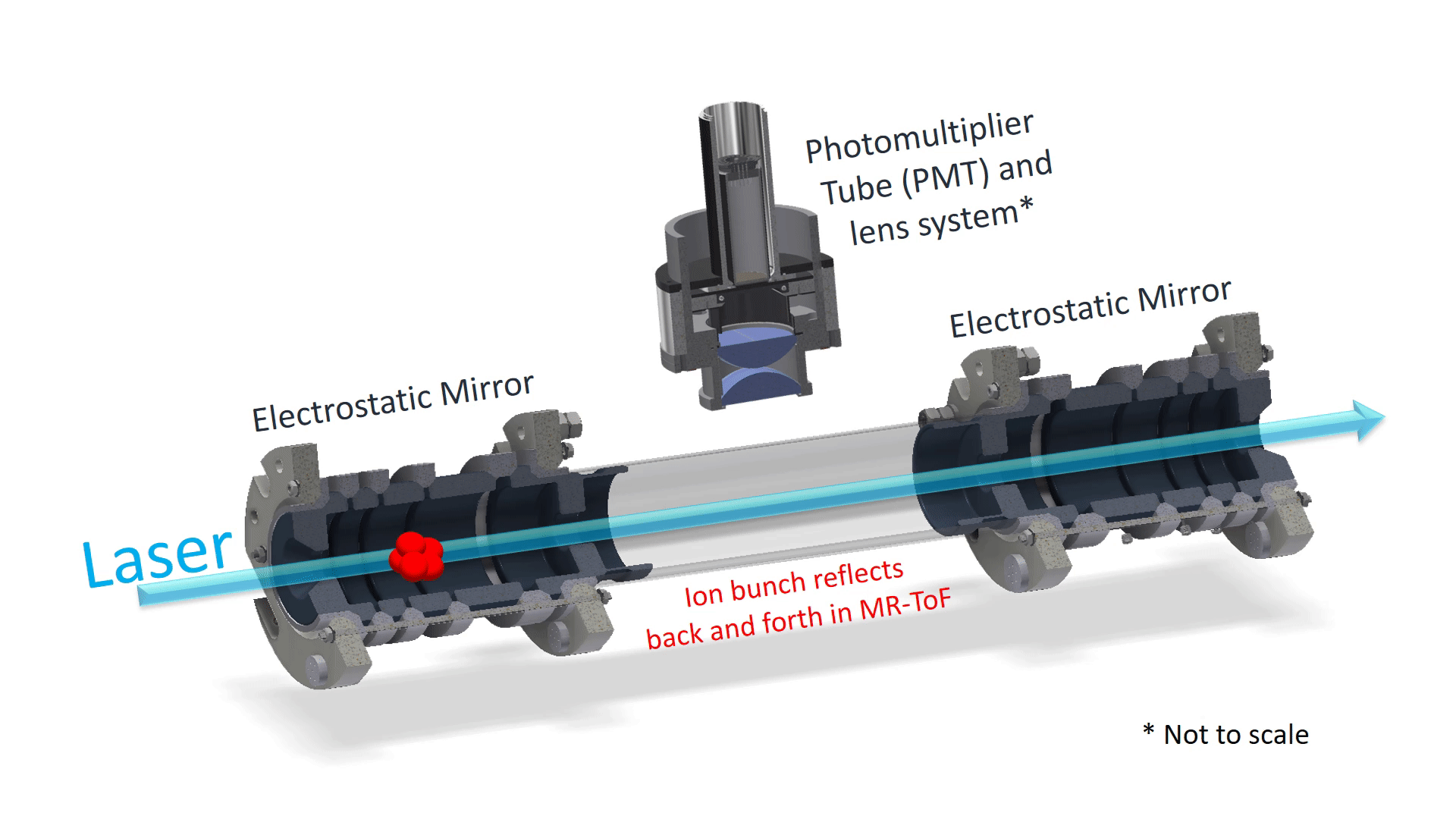
Fig 1: Drawing of the Multi-Reflection Time-of-Flight trap. The trapped ion bunch travels along the axis of the trap and laser. The photons emitted in the decay of the excited ions are detected by the photon detector located above the trap.
MIRACLS is an experiment located at the radioactive ion beam facility ISOLDE at CERN .
The name MIRACLS is an acronym that stands for Multi Ion Reflection Apparatus for Collinear Laser Spectroscopy. The goal of the experiment is to measure fundamental physical properties of exotic radioactive isotopes. These physical properties, such as the size, shape and electromagnetic moments of the isotopes are extracted from precise measurements of their hyperfine structure using laser spectroscopy. MIRACLS aims to increase the sensitivity of conventional fluorescence collinear laser spectroscopy (CLS) by trapping the probed ion bunches in a multi-reflection time-of-flight ion trap (MR-TOF). By doing so, the ion bunch that is only measured once in conventional collinear laser spectroscopy, can now be probed up to several ten thousands of times.
Currently, an MR-TOF operating at ion bunch energies of 30 keV is being designed, while an offline device is used to show the proof-of-principle and to both characterize and perfect the new technique. A schematic layout of the proof-of-principle setup is shown in Fig 2.
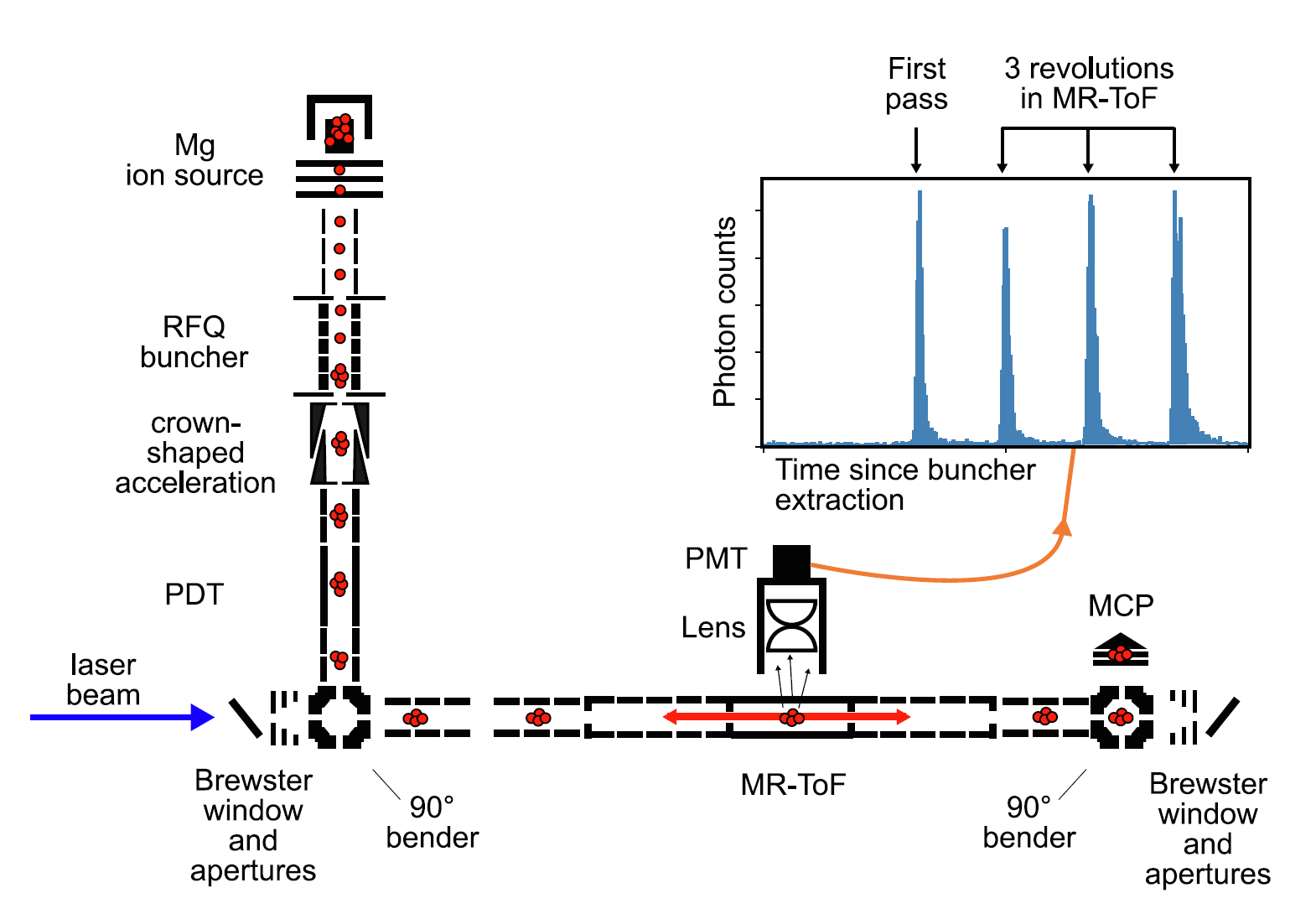
Fig 2: Layout of the proof-of-principle experiment at CERN. Ions, indicated by the red balls, are extracted from the ion source before they are cooled and bunched in the radio-frequency quadrupole (RFQ) buncher. The bunch is accelerated by a pulsed drift tube (PDT) and bent onto the axis of the MR-ToF and laser. Within the MR-ToF, the ion bunch is reflected back and forth between the electrostatic mirrors for several thousands of revolutions. The inset shows a photon signal as a function of time where every pulse indicated the ion bunch passing in front of the detector. After ejection from the trap, the ion bunch can be measured by a multi-channel plate detector (MCP).
In the proof-of-principle device, an ion source creates magnesium ions. These ions are sent towards a Radiofrequency quadrupole (RFQ) buncher. This device is a segmented linear Paul trap filled with helium buffer gas that is used to trap, cool and bunch the ions. As the ion bunches are released from the RFQ buncher, they are accelerated in the crown-shaped acceleration electrode and Pulse Drift Tube (PDT). A 90-degree bender sends the ion bunches onto the axis of the MR-ToF and laser beam. Upon entering the MR-ToF, the ion bunches are trapped between the two electrostatic mirrors of the device, where they perform several thousands of revolutions. With each revolution the ion bunch makes in the MR-ToF, the laser beam which is overlapped with their path, excites the ions resonantly. In the de-excitation process that follows, a photon is emitted by the ion. This photon is detected by a Photomultiplier tube (PMT) located above the MR-ToF device. This results in a time-structured photon signal that is recorded by the PMT as shown in the inset of the figure. In conventional fluorescence-based laser spectroscopy, only a single photon peak would be observed. In this figure, the signal is shown where only 3 revolutions in the MR-ToF were recorded using MIRACLS-type collinear laser spectroscopy. By scanning the laser frequency, the hyperfine structure can be measured. From this measurement, the size, shape and electromagnetic properties of the nucleus can be detected.